
Our new study on the biomechanics of Neanderthal’s anterior dentition has been recently published in the Journal of Human Evolution.
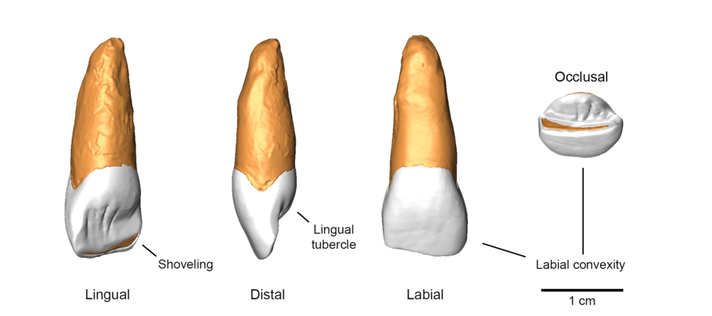
Neanderthal anterior teeth are very large (significantly larger than those of modern humans) and have a distinctive morphology characterised by robust “shovel-shaped” crowns. These features are seen as adaptive responses in dissipating heavy mechanical loads resulting from masticatory and non masticatory activities. This assumption (Anterior Dental Load Hypothesis; ADLH) is based on the heavily worn front teeth that characterise most Neanderthal specimens. The high frequency of enamel chipping, antemortem tooth loss and micro-fractures of the anterior teeth, have been considered as signals of extensive use of teeth as a third hand, in tearing, holding and shaping a variety of objects.
Although the long-standing debate surrounding this hypothesis has played a central role in paleoanthropology, is still unclear if Neanderthal anterior teeth can resist high mechanical loads or not. We answered this question by virtually simulating the chewing behaviour of the Neanderthal specimen of Le Moustier 1 to identify the occlusal contacts during edge-to-edge occlusion. We then used these contacts to load the finite element models of Le Moustier’s maxillary central incisor and compared the stress distribution with those of Qafzeh 9, an Early Homo sapiens from Israel dated to ca. 115–94 ka.
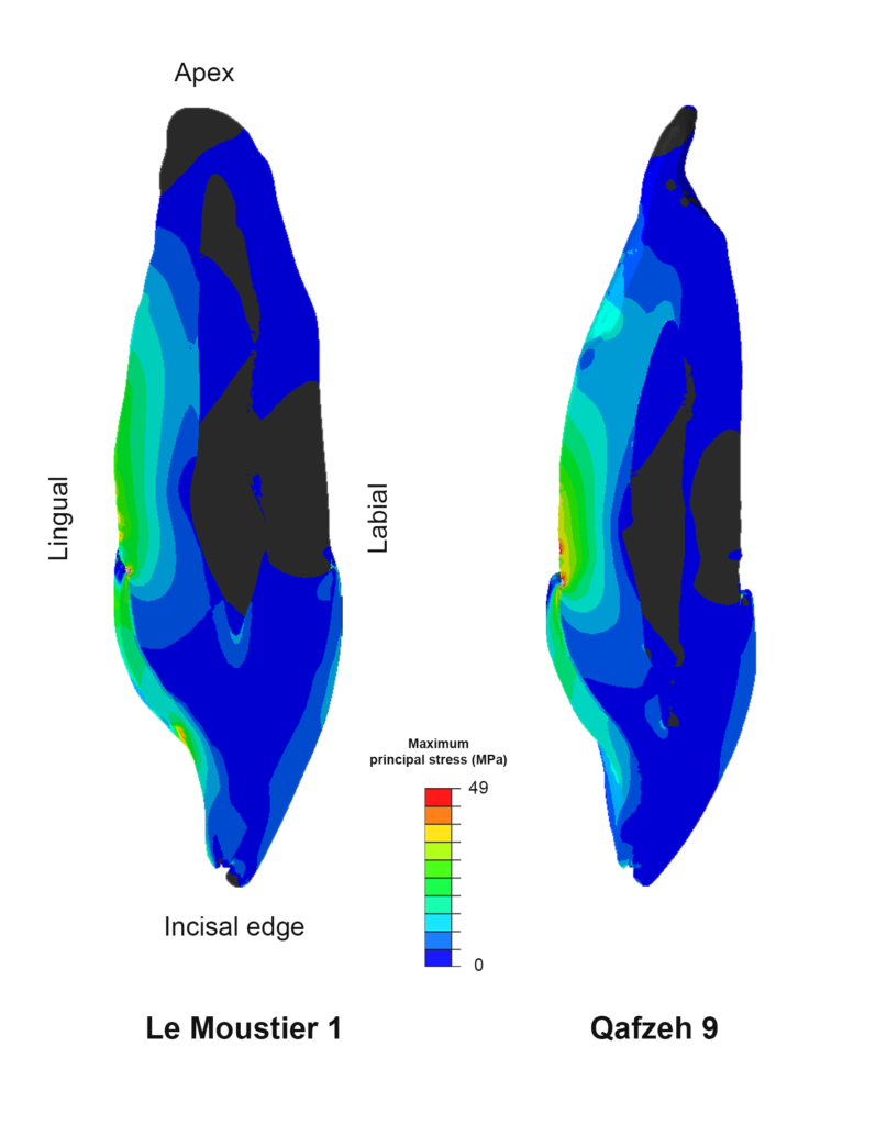
The results show stress distribution differences between Le Moustier 1 and Qafzeh 9, with the former displaying higher tensile stress in enamel around the lingual fossa but lower concentration of stress in the lingual aspect of the root surface. Moreover, we observed that the tensile stress in enamel is distributed lingually, while compressive stress develop labially. Our results also confirm that enamel-dentine junction represents an important intermediate tissue that helps in absorbing tensile stress. These results seem to suggest that the presence of labial convexity, lingual tubercle and of a large root surface in Le Moustier 1 incisor helps in dissipating mechanical stress. The absence of these dental features in Qafzeh 9 is probably compensated by the presence of a thicker enamel, which helps in reducing the stress in the tooth crown.